
Seated Hamstring Stretch
The seated hamstring stretch focuses on lengthening the muscles at the back of your thigh without putting unnecessary strain on your spine.
Osteoarthritis of the knee is the most common form of arthritis, caused by gradual wear and degeneration of the cartilage that cushions the knee joint. It is a leading cause of chronic joint pain and disability, particularly in older adults.

While knee osteoarthritis is most commonly associated with aging, it can also develop earlier in life due to injury, repetitive joint stress, or underlying joint conditions.
Understanding your condition is the first step toward management. Whether your goal is to stay active, reduce pain, or avoid surgery, this guide covers everything from anatomy to advanced physiotherapy rehabilitation.
The knee is one of the largest weight-bearing joints in the body, formed by the thigh bone (femur), shin bone (tibia), and kneecap (patella). Smooth articular cartilage covers the ends of these bones, allowing frictionless movement, while the meniscus acts as a shock absorber between the femur and tibia.
In osteoarthritis, this protective cartilage gradually breaks down. As cartilage thins and wears away, the underlying bone surfaces are exposed to increased friction and load, leading to pain, stiffness, and the formation of bony growths called osteophytes (bone spurs).
If you have knee osteoarthritis, you may notice the following signs:
Pain in the knee, especially during movement, weight-bearing, or after periods of activity.
Joint stiffness, particularly noticeable after waking up or sitting for prolonged periods.
Mild to moderate swelling around the knee joint due to inflammation.
Tenderness when pressing around the joint line.
Reduced ability to fully bend or straighten the knee.
Formation of small bony growths (osteophytes) around the joint margins.
A popping, cracking, or grinding sensation felt or heard during knee movement.
Knee osteoarthritis develops due to the progressive breakdown of joint cartilage. While aging and natural wear are the primary drivers, several factors can directly damage the joint and accelerate the condition.
Normal Wear and Tear: Cartilage naturally thins with age, reducing its ability to absorb shock.
Trauma or Injury: Falls, car accidents, or sports injuries that damage joint structures.
Repetitive Joint Stress: Occupational or recreational activities that place repeated load on the knee.
Inflammatory Conditions: Underlying inflammatory arthritis such as Rheumatoid Arthritis or Gout can damage joint surfaces and lead to secondary OA.
Certain factors may increase your likelihood of developing knee osteoarthritis:
Age: Adults older than 55 years are more prone to developing OA.
Gender: Women, especially during and after menopause, have a higher risk.
Obesity: Excess body weight significantly increases the load placed on the knee joint.
Genetics: A family history of osteoarthritis increases individual risk.
Diabetes: Associated with increased risk and faster progression of OA.
High Cholesterol: Linked to increased joint inflammation and cartilage degeneration.
Autoimmune Disease: Conditions affecting the joints can increase susceptibility to secondary OA.
If left unmanaged, knee osteoarthritis can lead to:
Chronic Pain: Persistent joint pain affecting daily function and quality of life.
Joint Stiffness: Progressive stiffness that limits mobility over time.
Psychological Impact: Depression and sleep disturbances may develop as a result of ongoing severe pain.
Reduced Mobility: Difficulty walking, climbing stairs, or performing daily activities as the condition progresses.
While osteoarthritis cannot always be prevented, especially with age, you can reduce your risk and slow its progression by maintaining good joint health through:
Avoiding Tobacco Products: Smoking is linked to faster cartilage degeneration.
Healthy Diet: Following a balanced, anti-inflammatory diet supports joint health.
Low-Impact Exercise: Regular low-impact activity helps maintain joint function without excessive strain.
Protective Equipment: Wearing appropriate protective gear during sports and physically demanding activities reduces injury risk.
Diagnosis of knee osteoarthritis is primarily made through physical examination and imaging tests.
Physical Examination
Your physiotherapist will assess joint swelling, tenderness, range of motion, alignment, and gait pattern, comparing both knees where applicable.
Imaging and Tests
X-Ray: Helpful to check for joint space narrowing and any calcium deposits within the joint.
MRI / CT: Used to identify other possible joint or bone problems not visible on X-ray.
Blood Tests: Used to rule out other conditions that cause similar symptoms, such as inflammatory or autoimmune arthritis.
There is no complete cure for knee osteoarthritis, as the underlying joint changes cannot be reversed. However, symptoms are completely manageable with the right combination of treatment.
Pain relievers such as topical creams and ointments can help manage localized pain and inflammation.
Use of braces, canes, or walkers to reduce joint load and improve stability during daily activities.
Reserved for severe cases that do not respond to conservative management. Arthroplasty (joint replacement) may be considered when the joint is significantly damaged.
Physiotherapy plays a central role in managing knee osteoarthritis, focusing on reducing pain, improving function, and slowing further joint deterioration. A combination of the following approaches is used based on your specific stage and severity.
Patient Education
Understanding your condition is key to long-term management. We help you understand the nature of OA, realistic expectations for recovery, and how to balance activity with joint protection in daily life.
Pain Management
Modalities: TENS and therapeutic Ultrasound (UST) help reduce pain signals and ease discomfort during flare-ups.
Heat & Cold Therapy: Heat therapy helps relax stiff muscles and improve blood flow before exercise, while cold therapy is useful after activity to reduce pain and inflammation.
Manual Therapy
Hands-on techniques such as joint mobilization and soft tissue release can help reduce stiffness, improve joint mechanics, and ease pain particularly useful when stiffness limits participation in exercise.
Range of Motion Exercises
Gentle, controlled movements through flexion and extension help maintain joint mobility, reduce stiffness, and prevent further loss of motion. These are usually introduced early and performed regularly, even during painful flare-ups.
Muscle Strengthening Exercises
Strengthening the muscles surrounding the knee particularly the quadriceps, hamstrings, and hip muscles is one of the most effective long-term strategies for reducing joint load and improving stability.
Early Stage: Gentle isometric exercises (e.g., quad sets) to activate muscles without stressing the joint.
Progressive Stage: Strengthening using light weights or resistance bands to build muscle support around the joint as tolerated.
Aerobic & Functional Exercise
Low-impact aerobic activity is essential for overall joint and general health:
Walking, Cycling, Swimming: Help reduce body weight, improve cardiovascular fitness, and increase blood circulation to the joint which in turn improves sleep quality and reduces pain perception.
Functional Training: Practicing daily movements like sit-to-stand, stair climbing, and walking patterns helps translate strength gains into real-world function.
Gait and Balance Training
Many individuals with knee OA develop compensatory walking patterns to avoid pain. Gait retraining and balance exercises help correct these patterns, reduce strain on the joint, and lower the risk of falls.
Weight Management Guidance
Since excess body weight significantly increases load on the knee joint, physiotherapists often work alongside guidance on activity-based weight management as part of a holistic treatment plan.
Long-Term Activity Planning
Physiotherapy isn’t just about symptom relief it’s about helping you stay active sustainably. We help design an ongoing exercise routine you can maintain independently to slow disease progression and preserve function over the long term.

The seated hamstring stretch focuses on lengthening the muscles at the back of your thigh without putting unnecessary strain on your spine.

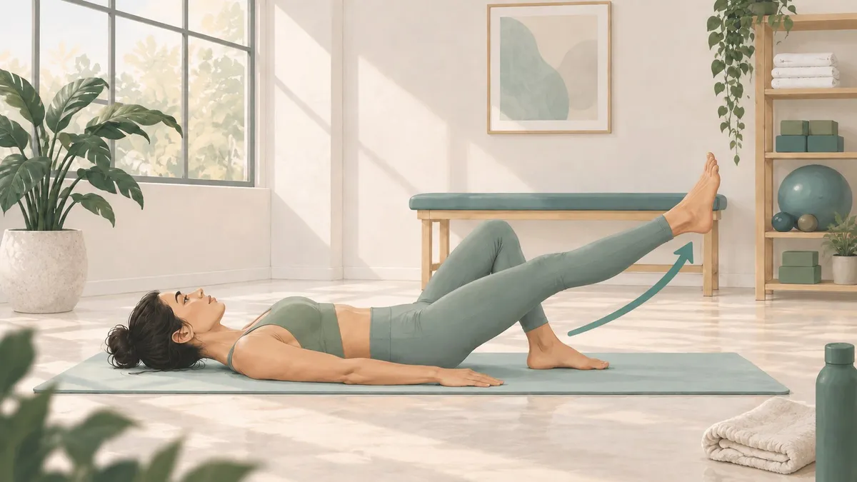
The Supine Straight Leg Raise (SLR) is a fundamental, closed chain like therapeutic exercise designed to isolate and strengthen the anterior thigh and hip musculature without placing excessive shear force on the knee joint.

Quadriceps isometrics are static exercises where you tighten your thigh muscle without actually moving your knee joint. It is a fantastic way to keep your leg strong when moving the joint hurts too much.

Hamstring isometrics in lying activates and strengthens the hamstring muscles using a towel for resistance without any knee movement, making it safe for early rehabilitation and pain free strengthening.
Book a consultation to get personalized treatment advice for your condition.
Book on Instagram